
研究ネットワークで、褐毛和種のかかえる問題解決を!
「高知大学 農林海洋科学部」 研究成果一覧
その他平成29年度生物系特定産業技術研究支援センター事業報告書
- 内容
- 平成29年度補正予算で実施した「革新的技術開発・緊急展開事業 (うち技術開発・成果普及等推進事業)」の事業成果報告書を公開いたします。
対象領域名「肉用牛 (褐毛和種) 生産」
担当者:松川 和嗣 (国立大学法人高知大学)
内容:
(1) 平成31年3月3日に開催したシンポジウムの概要
(2) 対象領域での生産者ニーズ
(3) 今後の技術開発・普及が必要な項目とその実現に向けた道筋を取りまとめた展望
- PDFファイルを閲覧する
学会発表凍結乾燥によるウシ体細胞の保存とその応用
- 発表者
- 松川 和嗣、本郷 新、田村 慎之介、枝重 圭祐 (高知大学農林海洋科学部)
- 学会名
- Cryopreservation Conference 2016
- 会場
- 基礎生物学研究所
- 開催日
- 平成28年11月10日~11日
詳細
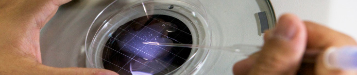
一般的に哺乳動物の体細胞は、超低温冷凍庫あるいは液体窒素中に”生きたまま“保存される。しかしながら、凍結細胞の維持や輸送にはコストや手間がかかる、液体窒素の取り扱いには安全面で問題があるなど欠点が認められる。さらに家畜では、災害や病疫等の緊急事態に備えての分散保存が望まれており、新たな保存技術が求められている。凍結乾燥 (フリーズドライ) 保存は減圧下での昇華を通じて水分を除去し保存する方法で、食品、医薬品、酵母等の長期保存のために実用化されているが、これまでに哺乳動物体細胞への応用例は少ない。高知県には褐毛和種高知系という独自に改良した和牛品種が存在し、近年その飼養頭数はわずか1,800頭となっている。我々は、希少な褐毛和種高知系の遺伝資源保存のためにクローン技術を介した体細胞の保存および個体再生に凍結乾燥技術が適用できるのではないかと考えた。そこで本研究では、ウシ凍結乾燥線維芽細胞の特性を調査し、凍結乾燥細胞の核移植後の発生能の検討をおこなった。
ウシ線維芽細胞は褐毛和種高知系の新生子耳片から樹立した。細胞をm-EGTA (50 mM EGTA, 100 mM Tris HCl, pH 8.2) に浮遊させ、緩慢凍結あるいは液体窒素による急速凍結後、乾燥した。凍結乾燥前後の細胞の生存性、形態、およびDNA損傷はCalcein-AM染色、電子顕微鏡観察、およびアルカリコメットアッセイによって評価した。凍結乾燥後の細胞をウシ除核未受精卵に注入し、核移植後の発生能を検討した。
凍結乾燥後の細胞はすべて死細胞であった。凍結乾燥後のDNA損傷細胞の割合は、緩慢凍結のほうが急速凍結よりも有意に低い値となった (14% vs. 24%, P<0.05)。電子顕微鏡による観察では、凍結乾燥細胞の細胞膜は著しく損傷を受けていたが、核膜の損傷はほとんど認められなかった。核移植後の卵割率および胚盤胞期胚発生率はそれぞれ50%および10%であった。以上より、凍結乾燥後のウシ線維芽細胞は生存していないものの、DNA損傷は少なく核移植後の発生が認められ、凍結乾燥保存が哺乳動物体細胞の遺伝資源保存技術として応用できる可能性が示唆された。今後は、凍結乾燥体細胞の長期常温保存およびクローン技術による個体作出を目指す予定である。
学会発表L-アスコルビン酸2リン酸の培地への添加がウシ体外受精胚の割球分離後の発生に及ぼす影響
- 発表者
- 田村慎之介,細川真実、河野葵、枝重圭祐、松川和嗣(高知大学農林海洋科学部)
- 学会名
- 日本繁殖生物学会第109回大会
- 会場
- 麻布大学
- 開催日
- 平成28年9月11日~15日
詳細
【目的】これまでに我々は、L-アスコルビン酸2リン酸 (AA-2P) の培地への添加が凍結融解後のウシ体外受精胚、および凍結融解胚盤胞の切断2分離後の生存率向上に効果があることを報告している (日本繁殖生物学会第105回つくば大会)。一方、高知県で飼養されている褐毛和種高知系は約1,700頭しかおらず、受精卵移植による増頭を推進しているが、より効率的な胚生産が必要とされている。そこで本研究では、褐毛和種高知系の効率的な増頭を目的として、AA-2Pの培地への添加がウシ体外受精胚の割球分離後の発生に及ぼす影響を検討した。【方法】食肉処理場由来の卵巣より採取したウシ卵母細胞を、エストラジオール17β、ピルビン酸、FSHおよび10%FCSを添加したTCM199培地内で22時間体外成熟培養をおこなった。体外成熟後、体外受精をおこない、体外受精28時間後に2細胞期胚をプロナーゼで透明帯を溶解し割球を分離した。その後BSA添加CR1aa培地に500 μM濃度の AA-2Pを加えた添加区および無添加区にそれぞれ分離した割球を導入し20時間培養した。培養ディッシュにはWell of the Well (WOW) を用いた。両区とも体外受精48時間後に5%FCS添加CR1aa培地に代え、その後6日間培養した。体外受精後8日目における胚盤胞発生率および総細胞数をAA-2P添加区および無添加区で比較した。【結果】AA-2P 添加区および無添加区で20時間培養した割球のそれぞれの胚盤胞発生率は62%および44%となり、AA-2P添加区では無添加区よりも胚盤胞発生率が向上した。胚盤胞の総細胞数は、AA-2P 添加区および無添加区でそれぞれ51.5±20.3個および41.0±21.1個となった。
学会発表Metabolomics of liver and skeletal muscle in Japanese Brown Cattle-Kochi after feeding yuzu peel
- 発表者
- Akane Iwasa1, Yukiko Iwamoto1, Yu Takenaka1, Mitsuharu Urabe1, Yasuo Takemura1, Shuji Sakamoto2, Takuma Higuchi2, Katsuji Morioka1, Kazumasa Kakibuchi3, Yutaka Ishida3, Kazutsugu Matsukawa1
1 Faculty of Agriculture and Marine Science, Kochi University
2 Science Research Center, Kochi Medical School, Kochi University
3 Shikoku Research Institute Inc.
- 学会名
- AAAP 17th Animal Science Congress
- 会場
- 九州産業大学
- 開催日
- 平成28年8月22日~25日
詳細
Yuzu (Citrus junos) is considered a health food in Japan, and the consumption is substantially increasing. However, after the juice is extracted from fruit, most of the peel is treated as waste. On the other hand, the number of Japanese Brown Cattle-Kochi, which is one domestic breed of Wagyu, has been reducing, therefore, the total number is only 1,700 at present. In this study, we evaluated the metabolomics of liver and skeletal muscle in Japanese Brown Cattle-Kochi after feeding yuzu peel, and examined whether yuzu peel altered the metabolism and added a high value to their beef. This experiments were conducted from September in 2015 to February in 2016. For analysis of metabolite in the liver, a total of 8 cattle (Yuzu feeding group: 4, non-feeding group: 4) were examined. Moreover, for analysis of metabolite in the skeletal muscle, 10 Japanese Brown Cattle (yuzu feeding group: 4, non-feeding group: 6), 3 Japanese Black Cattle, 3 beef from United States, and 3 beef from Australia were examined. Yuzu peel paste of 2.5% was added to feed for two weeks before slaughter. After slaughter, livers were collected immediately. Skeletal muscles were vacuum-packed and stored at 0°C for 20 days. The metabolites were analyzed by CE-TOFMS. In the liver, metabolites, which participate in deoxidization (e.g. glutathione, anserine, and carnosine), were increased in yuzu feeding group. In the skeletal muscle, there were huge differences of metabolites among four kinds of beef. The amounts of amino acid were higher in US and Australian beef than Wagyu beef. Yuzu feeding group contained more succnic acid, glutathione, and carnosine than another group. These results suggest that yuzu alter metabolism in the liver of Japanese Brown Cattle-Kochi, and add a high value to their beef.
学会発表Factors affecting DNA damage in bovine spermatozoa after freeze drying
- 発表者
- Kazutsugu Matsukawa1, Mana Nitta1, Toshinori Oikawa2,Shinouke Tamura1, Mami Hosokawa1, Aoi Kono1, Satoshi Akagi3, Akihiko Ichikawa4,
Keisuke Edashige1
1 Faculty of Agriculture, Kochi University
2 Miyagi Prefectural Livestock Experiment Station
3 Institute of Livestock and Grassland Science, NARO
4 Department of Mechatronics Engineering, Meijo University
- 学会名
- AAAP 17th Animal Science Congress
- 会場
- 九州産業大学
- 開催日
- 平成28年8月22日~25日
詳細
Recently, freeze-drying has been proposed as a novel method to preserve mammalian spermatozoa. If bovine spermatozoa are commonly stored by freeze-drying, it becomes an effective preservation method against great disasters. However, no calves have been obtained by FD spermatozoa, although blastocysts were produced (Keskintepe et al., 2002, Martins et al., 2007, Hara et al., 2014). In this study, factors affecting DNA damage in bovine freeze-dried (FD) spermatozoa were examined. Frozen spermatozoa were thawed and washed by Percoll density gradient centrifugation. Washed spermatozoa were incubated in BO supplemented with 5 mM dithiothreitol (DTT) or 8 mM glutathione (GSH) for 10 min at 38.5℃. Then, spermatozoa were suspended in two freeze drying solution, m-EGTA (50 mM EGTA, 100 mM Tris HCl, pH 8.2) and Na-EGTA (50 mM NaCl, 50 mM EGTA, 10 mM Tris HCl, pH 8.2), respectively. After freeze-drying, alkaline comet assay was used to assess DNA damage in spermatozoa. The FD spermatozoa were observed by Transmission Electron Microscopy (TEM). FD spermatozoa were also injected into matured oocytes. The rate of DNA-damaged spermatozoa was 0% in Na-EGTA and 2% in m-EGTA. The rate of DNA-damaged spermatozoa in GSH treatment was significantly lower than that in DTT treatment (0% vs. 25%, P<0.05). After freeze-drying, the nuclear membranes treated by DTT were destroyed, however, the nuclear membranes treated by GSH were intact according to TEM observation. The blastocyst formation rates in GSH treatment and DTT treatment were 8.5% and 0%, respectively. In conclusion, the DNA damage in bovine spermatozoa are suppressed after freeze-drying, when they are washed by Percoll density gradient centrifugation, incubated with GSH, and suspended in Na-EGTA. And GSH treatment for FD spermatozoa is effective to produce blastocysts.
学会発表Trial of nuclear transfer from bovine freeze-dried fibroblast cells
- 発表者
- Shinnosuke Tamura1, Satoshi Akagi2, Shin Hongo1, Akihiko Ichikawa3, Toshinori Oikawa4, Keisuke Edashige1, Pasqualino Loi5,Kazutsugu Matsukawa1
1 Faculty of Agriculture and Marine Science, Kochi University
2 Institute of Livestock and Grassland Science, NARO
3 Department of Mechatronics Engineering, Meijo University
4 Miyagi Prefectural Livestock Experiment Station
5 University of Teramo
- 学会名
- AAAP 17th Animal Science Congress
- 会場
- 九州産業大学
- 開催日
- 平成28年8月22日~25日
詳細
Freeze-dried (FD) somatic cells have been proposed as a new tool for producing cloned animals, since it could overcome disadvantages of the current cryopreservation method. Recently it was succeeded to develop into blastocysts by nuclear transfer (NT) using FD somatic cells in the sheep and pig. Furthermore, chimeric mice were produced by ES cell lines from nuclear transferred blastocysts of FD cells. We investigated the influence of freeze drying solution on DNA damage of bovine fibroblast cells after lyophilization, and the ability of in vitro development of oocytes after NT. In addition, OCT4 and IFN-tau expression of NT and IVF embryos were examined. Bovine fibroblast cells were obtained from ear skin tissues of a Japanese Brown Cattle-Kochi. The cells were suspended in m-EGTA (50 mM EGTA, 100 mM Tris HCl, pH 8.2) and Na-EGTA (50 mM NaCl, 50 mM EGTA, 10 mM Tris HCl, pH 8.2), and lyophilized for 11.5 h. The DNA damage of the FD cells was evaluated by alkaline comet assay. The rehydrated cells were injected into enucleated bovine oocytes. Activation treatment was accomplished by ionophore A23187 and 6-dimethylaminopyridine. NT embryos were cultured in CR1aa medium for 7 days. The DNA-damaged cells were significantly fewer in m-EGTA than in Na-EGTA (12% and 24%, p < 0.05). The cleavage rates of NT embryos in m-EGTA and Na-EGTA were 50% and 56%, and the blastocyst formation rates were 10% and 0%, respectively. No significant difference in OCT4 expression was observed between NT and IVF embryos. Whereas IFN-tau expression of NT embryos was lower than that of IVF embryos. These results suggest that m-EGTA is better than Na-EGTA for a freeze drying solution. Further studies are necessary in order to produce bovine clones from FD cells.